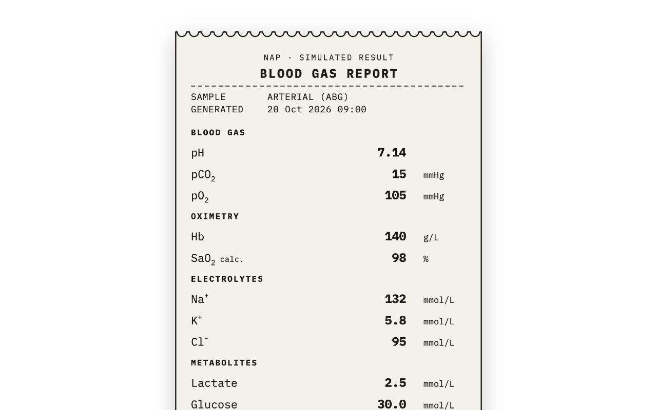
What it makes
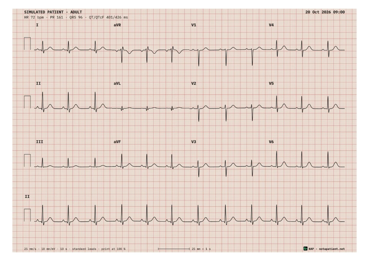
A ten-second, twelve-lead recording of a simulated patient, laid out the way a machine prints it.
- 3 × 4 plus a rhythm strip. I, aVR, V1, V4 / II, aVL, V2, V5 / III, aVF, V3, V6, then ten seconds of lead II.
- Calibrated. 25 mm/s, 10 mm/mV, a 1 mV pulse at the start of every row, a 1 mm and 5 mm grid.
- One heart, twelve views. Every lead comes from one model, so II = I + III and reciprocal changes appear where they should.
- Saved as a picture or a PDF. The PDF is vector and true scale on A4 or Letter; the picture is 300 dpi.
Rhythms and patterns
The 13 rhythms, rhythm settings and 12-lead patterns of the NAP controller's ECG tab, set with the controls above, by its rules.
- Rhythms. Sinus; AF, flutter, SVT and atrial tachycardia; 1° block, Mobitz I, Mobitz II and 3° block; VT, VF, torsades and asystole.
- STEMI by territory. Inferior, anteroseptal, high lateral, anterolateral, posterior and right ventricular, each with its reciprocal change.
- Conduction. RBBB, LBBB and WPW, which combine with a rhythm and a territory: RBBB with an inferior STEMI, WPW with pre-excited AF.
- Other patterns. Hyperkalaemia, pericarditis, LVH and Brugada type 1, plus ST depression and T inversion by territory.
- Rhythm settings. Occasional PVCs, Frequent PVCs, Bigeminy; coarse or fine VF; flutter conduction and atrial rate; the Mobitz conduction ratio; the 3° block escape rate; a QTcF target.
- Pacing. Fixed pacing at the age band's pacer rate, with or without capture.
- Chest leads. The standard V1–V6, posterior V7–V9 or right-sided V3R–V6R.
- Artefacts. None, or muscle tremor, baseline wander or both, the way a tense or shivering patient records.
Printing at true scale
Print the PDF at 100 % (“Actual size”, not “Fit to page”) and the sheet measures like ECG paper.
- Check with a ruler. The footer prints a 25 mm rule: one second, five large squares.
- 1 mV is 10 mm. At the standard gain the calibration pulse at the start of each row is two large squares tall.
- A4 or Letter. One layout, nothing scaled: the margins change, the millimetres do not.
Adult and paediatric
Choose an adult or one of four paediatric bands: infant, small child, bigger child or adolescent.
- Age-adjusted ECG morphology, grounded in published paediatric normal-limits data: axis, intervals, R/S progression and juvenile T waves change with the band.
- Teaching values. Plausible for teaching; not a validated paediatric reference.
How the ECG is made
One vector model per rhythm family drives all twelve leads: the limb leads are projections of a frontal-plane vector, the chest leads per-lead templates with territory-driven ST change.
Intervals, axes and amplitudes are informed by the sources listed on Evidence & methodology. It is a teaching ECG of a simulated patient: not a validated diagnostic recording, and never a real person.
Questions
- Is this a real patient’s ECG?
- No. Every sheet is generated from a model of a simulated patient, and its header says “Simulated patient”.
- Can I use the sheets in teaching materials?
- Yes. Use them as they are, with the NAP mark left on.
- Does it print at true scale?
- Yes, when the PDF is printed at 100 % (“Actual size”, not “Fit to page”). Check the 25 mm rule in the footer with a ruler: it is one second.
- Why is there no live view?
- The live 12-lead is inside a NAP session, where it follows the patient you control. This tool makes recordings to print and share.
- Can I fine-tune every parameter?
- This generator makes the patterns NAP teaches with, ready to print at true scale. To fine-tune literally any parameter, I recommend a dedicated ECG tool such as Squiggler, which displays virtually unlimited, realistic ECG variations.
In a NAP session
The same 12-lead opens on the learner's monitor and changes with the patient you control from your phone, with capture, gain and posterior or right-sided leads. Guide: 12-lead ECG